Using an algorithm they call the Krakencoder, researchers at Weill Cornell Medicine are a step closer to unraveling how the brain’s wiring supports the way we think and act. The study, published June 5 in Nature Methods, used imaging data from the Human Connectome Project to align neural activity with its underlying circuitry.
Mapping how the brain’s anatomical connections and activity patterns relate to behavior is crucial not only for understanding how the brain works generally but also for identifying biomarkers of disease, predicting outcomes in neurological disorders and designing personalized interventions.
Addressing the Elephant in the Room
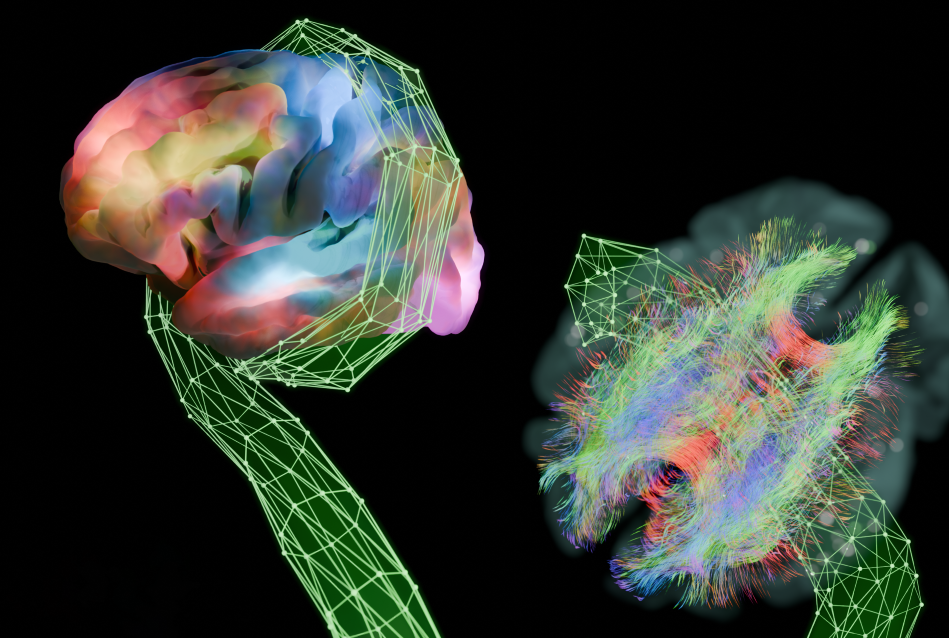
The brain consists of a complex network of interconnected neurons whose collective activity drives our behavior. The structural connectome represents the physical wiring of the brain, the map of how different regions are anatomically connected. The other piece of the puzzle is the functional connectome, which represents patterns of neuronal activity between different parts of the brain, highlighting regions that activate together during specific tasks or at rest. Surprisingly, scientists have found that regions that are “wired together” don’t always “fire together.”

Dr. Amy Kuceyeski
“But we’re still just scratching the surface of how brain networks relate to the tasks of everyday life, like solving a math problem, having a conversation with a friend or driving a car,” said senior author Dr. Amy Kuceyeski, professor of mathematics in radiology and in neuroscience the Feil Family Brain and Mind Research Institute at Weill Cornell Medicine.
While many researchers are modeling the relationship between functional and structural connectomes, they come up with different maps. “Everybody uses different methods to take pictures of the brain's networks,” Dr. Kuceyeski explained. For example, when using magnetic resonance imaging (MRI) for brain scans, different methods for processing the same raw images can generate different connectomes.
Dr. Kuceyeski likens this patchwork approach to examining an elephant in a dark room, where one person is touching the trunk, somebody a leg, someone else an ear. “Our fundamental assumption is that each set of choices in the imaging and processing pipelines provides a different view of the same underlying system,” said Keith Jamison, the study’s first author and a research associate in Dr. Kuceyeski’s lab.
To get a more comprehensive representation, Dr. Kuceyeski and her team built a tool that could take the structural and functional connectomes produced by all of these disparate approaches and collapse them together to produce a more unified interpretation.
“In my head, I saw it as some sort of monster with multiple arms that could reach out and grab different brain representations and digest and congeal them into one unified connectome,” Jamison said. The resulting program, an autoencoder that compresses and reconstructs more than a dozen different “flavors” of input data, thus came to be called the Krakencoder.
Identifying Connections that Restore Lost Function
The team trained the Krakencoder on data collected from over 700 subjects who participated in the National Institutes of Health’s Human Connectome Project. As part of that study, volunteers underwent extensive structural and functional MRI scanning.

Keith Jamison
The researchers found that the Krakencoder allowed them to take an individual’s structural connectome and correctly predict that person’s functional connectome about 20 times more accurately than previously published approaches.
The Krakencoder’s combined and compressed representation also predicted the individual’s age, sex and their cognitive performance scores received on tests which had been administered along with their imaging scans. Such scores, Dr. Kuceyeski noted, are notoriously difficult to gauge based on brain imaging alone.
Being able to map functions like cognition to specific brain networks is key to understanding how anatomy and physiology give rise to our behaviors and abilities—and how diseases and injuries can impair our performance.
In the future, Dr. Kuceyeski and her colleagues plan to combine the Krakencoder with a network modification tool they call NeMo that will allow them to examine the connectomes of people whose brains have been damaged by diseases. Christie Gillies, a PhD student in the Kuceyeski Lab, is using this approach to map outcomes following stroke.
“She's comparing the functional connectome produced by the NeMo + Krakencoder pipeline, based only on MRIs regularly collected in the clinic, to the actual functional MRI of a person. We found that our functional connectomes do a better job at predicting an individual’s motor scores and language scores at follow up,” Dr. Kuceyeski said.
These tools could also identify brain network connections associated with improved cognitive or motor performance. Boosting the activity of damaged circuits—for example, through transcranial magnetic stimulation, a treatment that uses magnetic pulses to stimulate nerve cells in the brain—could strengthen those connections and hasten recovery.
This research was supported by the National Institute of Mental Health grant RF1 MH123232, the Ann S. Bowers Foundation, the National Institute on Aging grant R01 AG053949 and the National Science Foundation CAREER 1748377.